
When your cardiologist recommends an angioplasty, you might hear them mention something called "IVUS" — a technology that allows them to see inside your arteries with remarkable precision. If this sounds like science fiction, you're not alone.
Here's the reassuring truth: IVUS (intravascular ultrasound) is one of the most significant advances in heart care over the past three decades. Research involving over 200,000 patients shows that IVUS-guided procedures lead to better outcomes.
In This Article
What Is Intravascular Ultrasound (IVUS)?
Intravascular ultrasound, or IVUS, is an imaging technique that uses sound waves to create detailed pictures of the inside of your coronary arteries — the blood vessels that supply your heart with oxygen and nutrients.
Think of It This Way
A regular angiogram (the X-ray movie of your heart arteries) shows you the shadow of your arteries from the outside, like looking at a tunnel from above. IVUS takes you inside the tunnel itself, showing you the walls, any buildup, and exactly how much space is left for blood to flow.
How Does IVUS Work?
During a coronary angiography or angioplasty procedure, your cardiologist can introduce a tiny ultrasound probe — about the thickness of a strand of spaghetti — into your coronary artery through the same catheter access point used for the main procedure.

Your cardiologist analyzes detailed IVUS images to make precise treatment decisions.
This miniature probe sends out sound waves that bounce off the artery walls and any plaque buildup. A computer then creates a detailed cross-sectional image showing:
True Artery Size
Often larger than what appears on angiogram
Plaque Amount & Type
Fatty, calcified, or mixed
Exact Narrowing
Precise measurements of blockage
Artery Wall Structure
Including hidden disease
Why Is IVUS Important?
The Limitations of Angiography Alone
Traditional angiography has been the gold standard for visualizing coronary arteries since the 1970s. It works well for identifying blockages, but it has limitations:
Shows only the silhouette — You see the channel where blood flows, but not the artery wall itself
Plaque can be hidden — Arteries can expand outward to accommodate plaque, making blockages appear less severe
Measurements are estimates — Sizing stents based on angiography alone requires some guesswork
Stent problems may be missed — Issues like incomplete expansion aren't always visible
What IVUS Reveals
IVUS overcomes these limitations by providing a complete 360-degree view of the artery:
See hidden plaque — Identifying disease that might otherwise be missed
Measure true artery size — Ensuring the right stent size is selected
Assess plaque composition — Including dangerous vulnerable plaques
Verify stent deployment — Confirming full expansion and proper positioning
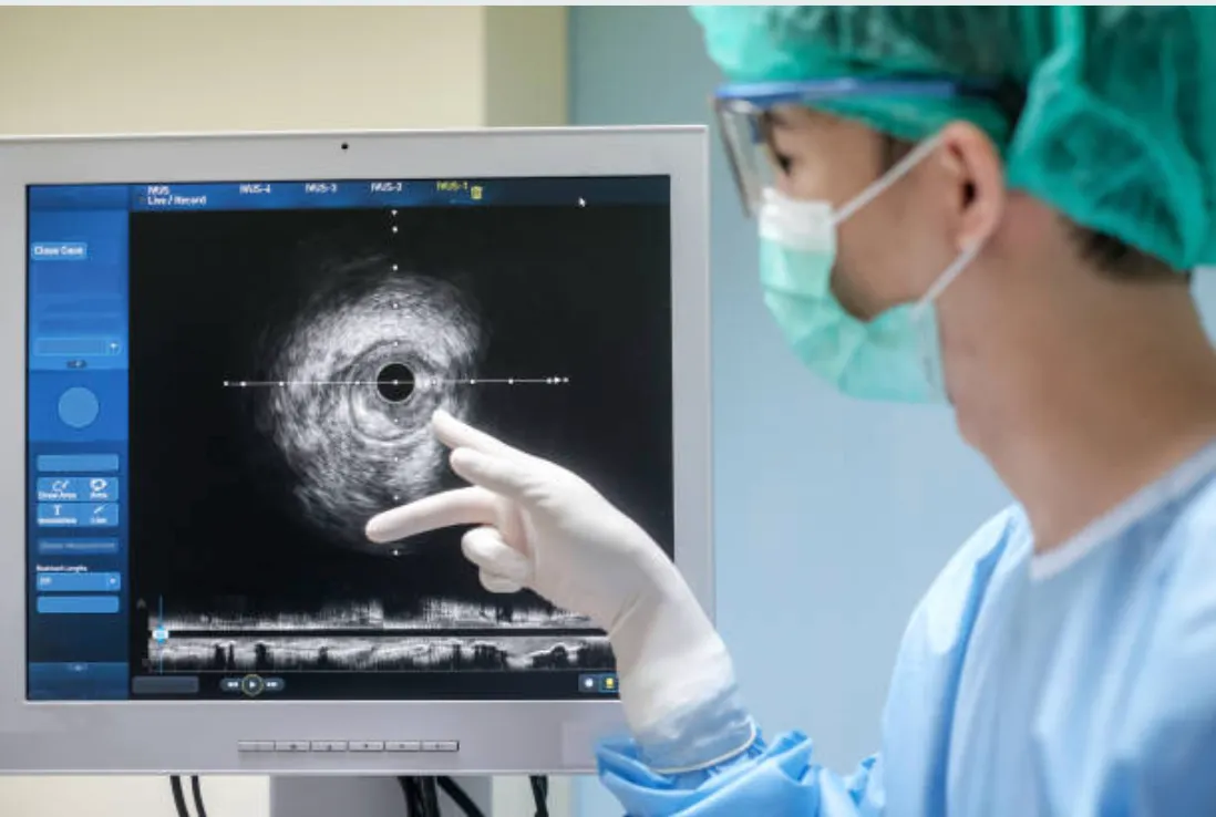
Real IVUS imaging: The doctor points to the cross-sectional view of the artery, showing the vessel wall and any plaque buildup in precise detail.
When Is IVUS Used?
IVUS is particularly valuable in certain situations:
Complex Blockages
- • Left main coronary artery disease — When the main trunk of your heart's blood supply is involved
- • Bifurcation lesions — Where arteries branch into two
- • Long, diffuse disease — When blockages extend over a significant length
- • Calcified arteries — Where hard calcium deposits make treatment challenging
Uncertain Findings
- • Intermediate blockages — When angiography shows 40-70% narrowing and it's unclear if treatment is needed
- • Hazy images — When the X-ray picture isn't definitive
- • Assessing previous stents — Checking for problems in stents placed earlier
Optimizing Results
- • Stent sizing — Choosing the correct diameter and length
- • Confirming expansion — Ensuring the stent is fully opened
- • Checking for complications — Identifying edge problems that need attention
The Evidence: Why IVUS-Guided Procedures Have Better Outcomes
The benefits of IVUS aren't just theoretical — they're backed by extensive research.
231,137 patients
Analyzed in a comprehensive meta-analysis of 52 studies — showing IVUS-guided procedures significantly outperform angiography-guided procedures.
Specific Outcomes from Research
A major analysis of nearly 28,000 patients found these benefits with IVUS guidance:
| Outcome | Reduction with IVUS |
|---|---|
| Cardiovascular death | 37% lower |
| Heart attack after procedure | 29% lower |
| Need for repeat procedure | 19% lower |
| Stent blood clots | 43% lower |
Even in Emergencies
A study of over 336,000 heart attack patients found that IVUS guidance was associated with 18% fewer adverse events compared to angiography guidance alone.
What to Expect: IVUS During Your Procedure

After your procedure, your cardiologist will explain what IVUS revealed and how it guided your treatment.
Before the Procedure
Your preparation is the same as for any angioplasty. There's nothing special you need to do because IVUS is being used.
During the Procedure
1. Procedure begins normally
Catheter access through your wrist or groin
2. Angiography performed
X-ray images identify the blockages
3. IVUS imaging
Ultrasound catheter advanced to area of interest
4. Pullback recording
Probe slowly withdrawn while recording detailed images
5. Treatment with guidance
Stents placed based on IVUS measurements
6. Confirmation
IVUS repeated to ensure optimal results
What You'll Feel
You won't feel the IVUS catheter itself — it's too small to cause sensation. The procedure may take slightly longer (15-30 minutes additional), but this extra time is invested in ensuring the best possible outcome.
Recovery is identical to any angioplasty. Most patients go home the same day or the next morning.
IVUS vs OCT: Two Ways to See Inside Arteries
You might also hear about OCT (optical coherence tomography), another way to image inside arteries:
| Feature | IVUS | OCT |
|---|---|---|
| Imaging method | Sound waves | Light waves |
| Image depth | Entire artery wall | Inner layers (high detail) |
| Resolution | Good (100-150 microns) | Excellent (10-15 microns) |
| Penetration | Deep — sees through plaque | Limited — blood must be cleared |
| Best for | Vessel sizing, plaque burden | Stent strut detail |
| Clinical experience | 30+ years | Newer technology |
Both technologies improve outcomes compared to angiography alone. Your cardiologist will choose based on your specific situation.
Frequently Asked Questions
Does IVUS add risk to the procedure? ▼
The additional risk from IVUS is minimal. The small ultrasound catheter is very safe, and the benefit of better-guided treatment far outweighs any tiny added risk. Major studies show that IVUS-guided procedures actually have better safety outcomes overall.
Will my insurance cover IVUS? ▼
IVUS is a well-established technology used routinely in cardiac catheterization labs worldwide. Coverage varies by insurance plan, but it's generally covered when medically indicated. Your hospital's billing team can provide specific information.
Does IVUS mean my case is more serious? ▼
Not necessarily. IVUS is used both for complex cases where it's clearly beneficial and for intermediate situations where more information helps with decision-making. Many cardiologists use IVUS routinely because it improves outcomes across a wide range of patients.
How long has IVUS been around? ▼
IVUS has been used clinically since the late 1980s — over three decades of experience. The technology has matured significantly, and there's extensive research supporting its benefits.
Will I be awake during IVUS imaging? ▼
Yes, just like a standard angioplasty. You'll receive sedation to help you relax and local anesthesia at the access site, but you'll be awake throughout. You won't feel the IVUS catheter moving through your arteries.
Does every patient need IVUS? ▼
While IVUS improves outcomes broadly, it's most beneficial in complex cases and intermediate lesions where additional information changes management. Your cardiologist will recommend IVUS when they believe it will benefit your care.
Is IVUS available at your hospitals? ▼
Yes, IVUS equipment is available at both our locations — Fortis Escorts Heart Institute, New Delhi and Preventia Clinic, Sector 76, Noida. Not every procedure requires it, but it's available when needed.
How does IVUS help with stent selection? ▼
IVUS provides precise measurements of your artery diameter and the length of diseased segments. This helps choose a stent that's the right size — not too small (which can lead to problems) and not too large (which can injure the artery).
Confidence from Clarity

Getting clear answers about your heart health empowers you to move forward with confidence.
When you're facing a heart procedure, it's natural to want the best possible care. IVUS represents one way that modern cardiology has moved beyond simple X-ray guidance to more sophisticated imaging that improves outcomes.
If you have questions about whether IVUS might be used in your procedure, ask your cardiologist. They can explain their approach and why certain imaging techniques are recommended for your specific situation.
References
- Şaylık F, et al. Comparison of Long-Term Outcomes Between IVUS-, OCT- and Angiography-Guided Stent Implantation: A Meta-Analysis. Angiology. 2024;75(9):809-819. PMID: 37644871
- Darmoch F, et al. IVUS-Guided Versus Angiography-Guided PCI: A Systematic Review and Meta-Analysis. J Am Heart Assoc. 2020;9(5):e013678. PMID: 32075491
- Kalsi J, et al. IVUS-Guided versus Angiography-Guided PCI for STEMI: Updated Meta-Analysis. Cardiology. 2024;149(3):196-204. PMID: 38350431
- Iuvara G, et al. Coronary Intravascular Imaging: A Comprehensive Review. Medicina (Kaunas). 2025;61(11). PMID: 41303855
- Khan SU, et al. IVUS-Guided PCI: Evidence and Practical Implications. Methodist Debakey Cardiovasc J. 2025;21(4):26-36. PMID: 40822366
- García-García HM, et al. High-definition IVUS: current clinical uses. Int J Cardiovasc Imaging. 2022;38(6):1213-1220. PMID: 38819587
- Sonoda S, et al. Current clinical use of IVUS to guide PCI. Cardiovasc Interv Ther. 2020;35(1):30-36. PMID: 31281937
About the Author
Dr. Shailesh Singh is a Consultant Interventional Cardiologist with over 12 years of experience in complex coronary interventions. He practices at Fortis Escorts Heart Institute, New Delhi and Preventia Clinic, Sector 76, Noida, where he performs IVUS-guided procedures for patients requiring advanced coronary imaging.
Learn more about Dr. Singh →Questions About Your Heart Procedure?
Dr. Shailesh Singh uses advanced imaging techniques including IVUS to ensure the best possible outcomes for his patients. Book a consultation to discuss your cardiac care.
Locations: Fortis Escorts Heart Institute, New Delhi | Preventia Clinic, Sector 76, Noida
Medical Disclaimer: This article is for informational purposes only and does not replace professional medical advice. Please consult with your cardiologist about your specific condition and treatment options.